| 图片: | |
|---|---|
| 名称: | |
| 描述: | |
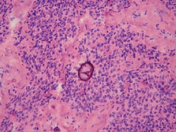
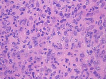
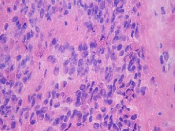
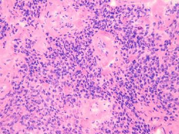
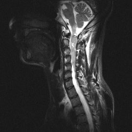
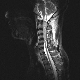
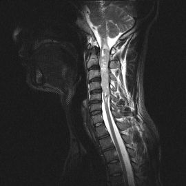
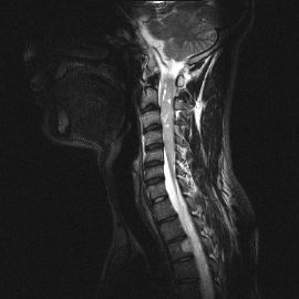
- 脊髓肿物
-
redsnow007 离线
- 帖子:329
- 粉蓝豆:5
- 经验:999
- 注册时间:2008-01-01
- 加关注 | 发消息
-
lanyueliang 离线
- 帖子:679
- 粉蓝豆:11
- 经验:1424
- 注册时间:2008-11-12
- 加关注 | 发消息
-
redsnow007 离线
- 帖子:329
- 粉蓝豆:5
- 经验:999
- 注册时间:2008-01-01
- 加关注 | 发消息
-
redsnow007 离线
- 帖子:329
- 粉蓝豆:5
- 经验:999
- 注册时间:2008-01-01
- 加关注 | 发消息
-
stevenshen 离线
- 帖子:343
- 粉蓝豆:2
- 经验:343
- 注册时间:2008-06-03
- 加关注 | 发消息
-
redsnow007 离线
- 帖子:329
- 粉蓝豆:5
- 经验:999
- 注册时间:2008-01-01
- 加关注 | 发消息
-
redsnow007 离线
- 帖子:329
- 粉蓝豆:5
- 经验:999
- 注册时间:2008-01-01
- 加关注 | 发消息
-
redsnow007 离线
- 帖子:329
- 粉蓝豆:5
- 经验:999
- 注册时间:2008-01-01
- 加关注 | 发消息

Diffuse craniospinal metastases of intraventricular rhabdoid papillary meningioma with glial fibrillary acidic protein expression: a case report.
Eom KS, Kim DW, Kim TY.
Department of Neurosurgery, Wonkwang University School of Medicine, Iksan 570-749, Republic of Korea.
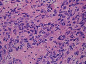
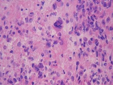
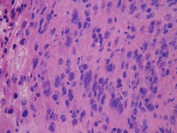
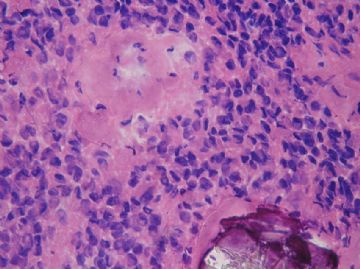
Rhabdoid papillary meningioma is a recently described clinically aggressive variant of meningiomas with a high recurrence rate. Additionally, only one case of intraventricular rhabdoid meningioma has been reported so far. We present a case of a 50-year-old man who developed an intracranial tumor of the left lateral ventricle at the trigone, for which he underwent total tumor resection followed by gamma knife radiosurgery for recurrence of the tumor. The histological diagnosis was rhabdoid papillary meningioma. Five years after surgery, diffuse craniospinal leptomeningeal metastases developed and subtotal removal of the spinal tumor was performed. The spinal tumor was considered to have metastasized via cerebrospinal fluid (CSF) in view of its histological features that were identical to those of the primary tumor. Immunohistochemistry revealed the unusual cytoplasmic expression of glial fibrillary acidic protein (GFAP) of tumor cells. To our knowledge, this is the first reported case of diffuse craniospinal metastases of intraventricular rhabdoid papillary meningioma with GFAP expression and the second reported case of the rhabdoid subtype amongst intraventricular meningiomas.
Publication Types:
PMID: 19482417 [PubMed - in process]-
2: Diagn Cytopathol. 2003 May;28(5):274-7.

Meningioma with extensive noncalcifying collagenous whorls and glial fibrillary acidic protein expression: new variant of meningioma diagnosed by smear preparation.
Pope LZ, Tatsui CE, Moro MS, Neto AC, Bleggi-Torres LF.
Department of Pathology (Neuropathology), Universidade Federal do Paraná, Curitiba, Brasil.
The purpose of this report is to describe the unique cytological findings of a new recently characterized type of meningioma that has extensive noncalcifying collagenous whorls and glial fibrillary acid protein (GFAP) expression. This new entity, described by Haberler and colleagues, was named whorling sclerosing variant of meningioma. The patient was a 34-yr-old white man with a large tumor in the brainstem. Intraoperative smear preparations showed a tumor with a large number of solid hyaline masses in a loose background and in focal areas tumor cells formed cohesive nests with a somewhat whorling appearance. The histological sections showed a neoplasia composed of innumerable eosinophilic, collagenous, noncalcified round deposits, cuffed by scattered meningothelial tumor cells. The neoplastic cells showed diffuse cytoplasmic reactivity for EMA and vimentin, as well as positivity to GFAP. This is the first cytological description of this new entity in the literature. Copyright 2003 Wiley-Liss, Inc.
Publication Types:
PMID: 12722124 [PubMed - indexed for MEDLINE]-
3: Acta Neuropathol. 1997 Nov;94(5):499-503.
An unusual meningioma variant with glial fibrillary acidic protein expression.
Su M, Ono K, Tanaka R, Takahashi H.
Department of Pathology, Niigata University, Japan.
We studied a recurrent meningioma located in the right frontal lobe. The tumor showed high cellularity and the cells had plump, hyalinous cytoplasm. Immunohistochemically, almost all the tumor cells were positive for epithelial membrane antigen and vimentin, and unexpectedly, glial fibrillary acidic protein (GFAP). Ultrastructural investigation revealed abundant 8- to 10-nm filaments in the cytoplasm. Conspicuous interdigitations with numerous desmosomes were present. Frequently, intracellular and intercellular lumina lined by microvilli were also found. We considered the present case to be an unusual variant of meningioma with GFAP expression. A few cases of meningioma with triple expression of GFAP, vimentin and cytokeratin have been reported previously. However, the present case showed obvious pathological differences from these, and had no immunoreactivity for cytokeratin.
Publication Types:
PMID: 9386784 [PubMed - indexed for MEDLINE]-
4: Acta Neuropathol. 1995;90(5):539-44.
Suprasellar meningioma with expression of glial fibrillary acidic protein: a peculiar variant.
Wanschitz J, Schmidbauer M, Maier H, Rössler K, Vorkapic P, Budka H.
Institute of Neurology, University of Vienna, Austria.
A 24-year-old female presented with a 3-year history of a suprasellar and intraventricular solid midline process measuring about 3 x 4 cm. At surgery, this tumour was sharply delineated and of stone-like firmness and was removed completely. Histology suggested meningioma, featuring nests and cords of epithelium-like cells with prominent cytoplasm amidst abundant fibrous stroma with prominent lymphoplasmocellular infiltration. Immunocytochemically, the tumour cells expressed vimentin, S-100 protein, epithelial membrane antigen, cytokeratins, and most surprisingly, glial fibrillary acidic protein (GFAP). Ultrastructural investigation revealed abundant intermediate filaments and occasionally dense secretory granules in tumour cells with short, finger-like cytoplasmic processes joined by very rare small, but well-developed desmosomes. This tumour most likely represents a peculiar variant of meningioma with prominent production of GFAP, as previously described [Budka H (1986) Acta Neuropathol (Berl) 72: 43-54].
Publication Types:
PMID: 8560989 [PubMed - indexed for MEDLINE]-
5: Acta Neuropathol. 1986;72(1):43-54.
Non-glial specificities of immunocytochemistry for the glial fibrillary acidic protein (GFAP). Triple expression of GFAP, vimentin and cytokeratins in papillary meningioma and metastasizing renal carcinoma.
Budka H.
In an extensive immunocytochemistry study for glial fibrillary acidic protein (GFAP) of human neuropathological biopsy or autopsy tissue specimens examined for diagnostic or research purposes, rare non-glial specificities of the GFAP immunostain were observed: Schwann cells of some small nerves in salivary gland, renal capsule, and in epidural fat adjacent to a metastatic carcinoma, Schwann and satellite cells in a spinal ganglion invaded by tumor, chondrocytes of epiglottic cartilage, few cells of a malignant pleomorphic adenoma of salivary gland, most cells of a recurrent papillary meningioma with areas similar to the hemangiopericytic variant, and many cells of a renal carcinoma metastatic to brain; the primary renal tumor had been operated 4 years earlier and focally contained some GFAP-positive cells. To ascertain the specificity of such unexpected immunoreactivities for GFAP and to exclude possible crossreactivities with other intermediate filament (IF) proteins, a panel of different antibodies was used for immunocytochemistry with the peroxidase-antiperoxidase (polyclonal antisera) or labeled biotin-avidin (monoclonal antibodies) techniques: two monoclonal and four polyclonal anti-GFAP, three monoclonal and one polyclonal anti-cytokeratins (CK), and two monoclonal anti-vimentin (VIM) antibodies. Triple expression of GFAP, VIM and CK was found in the papillary meningioma (in patterns suggesting frequent co-localization), in the metastatic carcinoma (in patterns suggesting little co-localization), and in the pleomorphic adenoma (only few GFAP-positive cells). Co-expression of GFAP and VIM was seen in epiglottic chondrocytes and reactive astroglia; another metastatic carcinoma was labeled only for CKs. In the light of previous reports on non-glial specificities of the GFAP immunostain, and of the consistency of our immunostaining results obtained by all monospecific anti-GFAP antibodies as well as the lack of immunocytochemically evident crossreactivity with other IF proteins, authentic GFAP production by some rare non-glial tissues and tumors is suggested.
请见下列文献:
1: Clin Neurol Neurosurg. 2009 Sep;111(7):619-23. Epub 2009 May 30.
Related Articles,
onmouseout="PopUpMenu2_Hide();" href="javascript:PopUpMenu2_Set(Menu19482417);" target="_self">Links

- 王军臣
